Coloring Worksheet Analysis

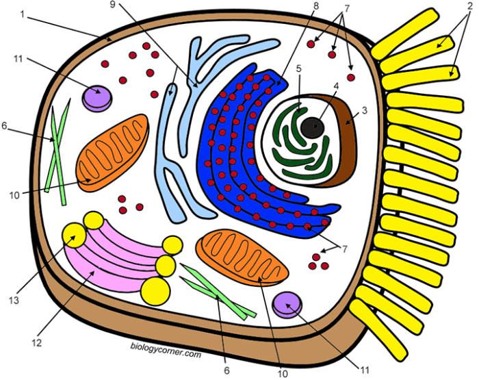
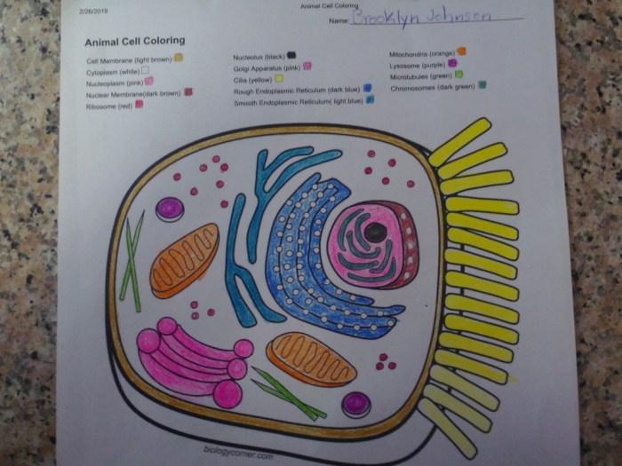
Animal cell coloring answer sheet – Welcome! Let’s delve into the fascinating world of the animal cell by examining the processes that make it tick. This analysis will focus on key cellular activities and how they relate to the specific organelles you’ve colored in your worksheet. Understanding these processes will give you a deeper appreciation for the intricate workings of even the smallest living unit.
Cellular processes are complex, coordinated events that occur within the cell to maintain life. Two vital processes are cellular respiration and protein synthesis, both reliant on the precise interactions between various organelles. The nucleus, the control center, orchestrates these activities.
So you’ve finished your animal cell coloring answer sheet? Great! Now, for a bit of a change of pace, why not check out some awesome african animal coloring pictures for a fun break? It’s a good way to relax after the precision work of the cell diagram, and then you can get back to focusing on those organelles!
Cellular Respiration and Protein Synthesis
Cellular respiration, the process of generating energy (ATP) from glucose, primarily occurs in the mitochondria. These organelles, often called the “powerhouses” of the cell, have a folded inner membrane (cristae) that increases surface area for the numerous enzyme-driven reactions involved in respiration. The glucose is broken down through a series of steps, ultimately producing ATP, the cell’s usable energy currency.
Protein synthesis, on the other hand, involves the creation of proteins from amino acids. This process begins in the nucleus, where the DNA contains the genetic code for protein structure. This code is transcribed into messenger RNA (mRNA), which then travels to the ribosomes. Ribosomes, located on the rough endoplasmic reticulum (RER) or free-floating in the cytoplasm, translate the mRNA code into a specific amino acid sequence, building the protein.
The Nucleus: The Cell’s Control Center
The nucleus houses the cell’s genetic material, DNA, organized into chromosomes. It acts as the control center, dictating all cellular activities by regulating gene expression. The DNA contains the instructions for making all the proteins the cell needs, which determine the cell’s structure and function. The nucleus controls which genes are “turned on” or “off,” determining which proteins are synthesized at any given time, thus influencing the cell’s response to its environment and its overall activity.
For example, a muscle cell will express genes related to muscle contraction, while a nerve cell will express genes related to signal transmission. This precise regulation ensures the cell functions correctly.
Endoplasmic Reticulum and Golgi Apparatus Collaboration
The endoplasmic reticulum (ER) and the Golgi apparatus work in concert to modify, package, and transport proteins. The rough ER, studded with ribosomes, synthesizes proteins. These proteins then move into the lumen of the ER, where they may undergo initial folding and modification. From the ER, proteins are transported to the Golgi apparatus, a stack of flattened sacs.
Here, proteins undergo further processing, sorting, and packaging into vesicles for transport to their final destinations within or outside the cell. Imagine the ER as a manufacturing plant and the Golgi apparatus as the shipping and packaging department; each plays a crucial role in the efficient delivery of cellular products. For instance, proteins destined for secretion are packaged into secretory vesicles that fuse with the plasma membrane, releasing the proteins outside the cell.
Creating an Enhanced Coloring Worksheet

Let’s elevate our animal cell coloring worksheet to a more engaging and informative experience for students. By adding complexity and detail, we can foster a deeper understanding of the intricate workings of an animal cell. This enhanced worksheet will not only be a fun activity but also a valuable learning tool.This section details the design and implementation of a more comprehensive animal cell coloring worksheet, including a legend and a section for independent exploration and drawing.
The goal is to create a worksheet that encourages active learning and reinforces key concepts.
Worksheet Design and Organization
The redesigned worksheet will feature a larger, more detailed illustration of an animal cell. Instead of simply outlining organelles, the drawing will show a more realistic representation, including the relative sizes and positions of different organelles within the cell. The level of detail will be appropriate for the age group, avoiding overwhelming complexity while still providing a stimulating challenge.
For example, the endoplasmic reticulum could be depicted as a network of interconnected tubules and sacs, rather than just a simple shape. Similarly, the Golgi apparatus could be shown as a stack of flattened sacs, highlighting its layered structure. The mitochondria could be illustrated with their inner and outer membranes, showing the cristae folds. The nucleus would be shown with a clearly defined nuclear envelope and nucleolus.
Legend and Color Coding, Animal cell coloring answer sheet
A comprehensive legend will be included, associating each organelle with a specific color. This will aid in both coloring and identification. For instance:
- Cell Membrane: Blue
- Cytoplasm: Light Yellow
- Nucleus: Purple
- Nucleolus: Dark Purple
- Rough Endoplasmic Reticulum: Light Green
- Smooth Endoplasmic Reticulum: Dark Green
- Ribosomes: Small Dark Brown Dots
- Golgi Apparatus: Orange
- Mitochondria: Red
- Lysosomes: Dark Blue
- Centrioles: Pink
This color-coding system will create a visually appealing and easily understandable guide for students. The choice of colors is intentional, aiming for clarity and avoiding colors that might be difficult to distinguish. The legend will be placed prominently on the worksheet, readily accessible for reference throughout the coloring process.
Section for Additional Cellular Structures
To encourage further exploration and critical thinking, a dedicated section will be provided for students to draw and label additional cellular structures not included in the main illustration. This section could include prompts such as: “Draw and label a vacuole,” or “Draw and label the cytoskeleton.” This open-ended section allows for creativity and deeper engagement with the material, allowing students to expand their knowledge beyond the basic organelles.
It also provides an opportunity for differentiation, allowing students to delve deeper into specific areas of interest. This section would encourage independent research and reinforce learning through active participation.
Helpful Answers: Animal Cell Coloring Answer Sheet
What are some common mistakes students make when using an animal cell coloring answer sheet?
Common mistakes include inaccurate labeling of organelles, misinterpreting the size and shape of organelles, and failing to connect organelle structure to function.
How can an animal cell coloring answer sheet be adapted for different age groups?
Simpler worksheets with fewer organelles can be used for younger students, while more complex worksheets with additional details and processes can be used for older students. The level of detail in the accompanying descriptions should also be adjusted accordingly.
Are there online resources available to help students create their own animal cell coloring answer sheets?
Yes, numerous online resources provide templates, diagrams, and information about animal cell structures, enabling students to create personalized worksheets.
