Developing Coloring Activities

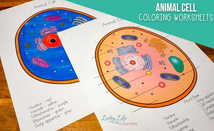
Animal cell worksheet coloring – This section details three distinct coloring activities designed to engage students with different aspects of animal cell structure, catering to varying skill levels. Each activity focuses on a specific organelle and employs different coloring techniques to enhance learning and creativity. The activities are designed to be presented in a sequential order, progressing from simple to complex.
Nucleus Coloring Activity: Simple
This activity focuses on the basic structure of the nucleus. Students will color a simplified representation of the nucleus, including the nuclear envelope and nucleolus. The coloring technique is straightforward, using a single color for the nucleus and a slightly different shade for the nucleolus to differentiate the structures. For example, the nucleus could be colored a light purple, while the nucleolus could be a darker shade of purple.
This simple activity introduces the concept of the nucleus as the control center of the cell.
Mitochondria Coloring Activity: Medium Complexity, Animal cell worksheet coloring
This activity introduces the concept of the mitochondria’s internal structure. Students will color a more detailed representation of a mitochondrion, including the inner and outer membranes, cristae, and matrix. The coloring technique involves using multiple colors to differentiate the different parts of the organelle. For instance, the outer membrane could be a light blue, the inner membrane a darker blue, and the cristae a reddish-brown.
The matrix could be a light yellow. This activity reinforces the understanding of the mitochondria’s role in energy production.
Cell Membrane Coloring Activity: Advanced Complexity
This activity focuses on the complex structure and function of the cell membrane. Students will color a representation of the cell membrane, including the phospholipid bilayer, embedded proteins, and cholesterol molecules. The coloring technique will involve a detailed representation of the membrane’s structure, using various colors and patterns to represent the different components. For example, the phospholipid heads could be colored red and the tails blue, with different colored shapes representing the proteins and cholesterol molecules.
This activity promotes a deeper understanding of the cell membrane’s selective permeability and its role in cell communication.
Coloring Techniques: Simple, Medium, and Advanced
The coloring activities utilize a progression of coloring techniques to enhance engagement and understanding. Simple techniques involve single-color filling of shapes. Medium techniques incorporate multiple colors to distinguish different parts of an organelle. Advanced techniques utilize shading, texture, and patterns to represent the complex structure and function of the cell membrane, allowing students to explore the finer details of its composition.
Students might, for example, use a stippling technique to represent the protein density within the cell membrane.
Animal cell worksheet coloring offers a unique blend of education and fun, allowing students to visualize complex biological structures. For a change of pace, consider supplementing this activity with some delightful illustrations, perhaps using the charming farm animal coloring pages to reinforce the concept of diverse cell types found in various organisms. Returning to the animal cell worksheet, remember to carefully color each organelle to fully grasp its function within the cell.
Worksheet Design Sequence
The worksheet is designed with a logical progression of complexity. The nucleus activity, being the simplest, is presented first, followed by the mitochondria activity of medium complexity, and finally, the cell membrane activity, which is the most complex. This sequence allows students to gradually build their understanding of animal cell structures and coloring techniques.
Creating Engaging Visuals for the Worksheet: Animal Cell Worksheet Coloring

A visually appealing worksheet is crucial for maintaining children’s interest and facilitating effective learning. The color scheme and illustrations should be carefully chosen to not only be aesthetically pleasing but also to aid in understanding the complex structures and functions of an animal cell.Color palettes should be thoughtfully selected to enhance comprehension and avoid overwhelming the viewer. Using a range of colors improves clarity and allows for easy differentiation between organelles.
Color Scheme Selection
A vibrant yet scientifically appropriate color scheme is essential. We propose a palette incorporating muted greens and blues for the cytoplasm and cell membrane, representing their natural, organic qualities. These calming colors provide a neutral backdrop to highlight the organelles. Bright, contrasting colors like oranges, yellows, and pinks can be used to depict the nucleus, mitochondria, and Golgi apparatus, respectively, emphasizing their distinct roles in cellular processes.
Purples and reds can effectively represent lysosomes and vacuoles, conveying their functional importance. This contrast helps children quickly identify and associate colors with specific organelles.
Animal Cell Illustrations
The effectiveness of the worksheet hinges on clear and engaging illustrations. Different levels of detail cater to varying age groups and learning levels.
Below are descriptions of three animal cell illustrations, ranging in complexity:
Simple Animal Cell Illustration
This illustration presents a highly simplified representation of an animal cell. The cell is depicted as a circle with a large, centrally located nucleus (yellow) and a smaller, oval-shaped mitochondria (orange) near the nucleus. The cytoplasm (light green) fills the remaining space, and the cell membrane (darker green) forms the outer boundary. This simplistic representation focuses on the essential components, making it ideal for younger learners.
The color contrast between the nucleus and mitochondria and the cytoplasm is stark to emphasize their individual identities.
Moderately Detailed Animal Cell Illustration
This illustration builds upon the simple version by adding more organelles. The nucleus (yellow) retains its central position, but now includes a visible nucleolus (darker yellow). The mitochondria (orange) are depicted as multiple bean-shaped structures scattered throughout the cytoplasm (light green). The Golgi apparatus (pink) is shown as a stack of flattened sacs near the nucleus. A few lysosomes (purple) are scattered within the cytoplasm.
The cell membrane (dark green) remains a distinct outer boundary. This level of detail introduces more complexity while remaining easily digestible for older elementary school children. The use of different shades of yellow within the nucleus aids in distinguishing the nucleolus.
Highly Detailed Animal Cell Illustration
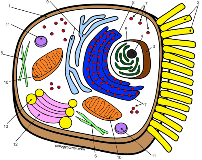
This illustration provides a comprehensive representation of an animal cell, including a detailed nucleus (yellow) with a clearly defined nucleolus (darker yellow) and chromatin (light yellow). Numerous mitochondria (orange) are shown, varying in size and shape to reflect their dynamic nature. The endoplasmic reticulum (light blue), both rough and smooth, is depicted as an extensive network of interconnected membranes.
The Golgi apparatus (pink) is shown as a more complex structure with numerous vesicles budding off. Lysosomes (purple) and vacuoles (light red) are more numerous and varied in size. Ribosomes (small dark grey dots) are scattered throughout the cytoplasm (light green) and on the rough endoplasmic reticulum. The cell membrane (dark green) is shown with embedded proteins (small, differently colored shapes).
This illustration is suitable for older students and provides a detailed overview of the cell’s intricate structure. The diversity in shapes and sizes of organelles within the cytoplasm aids in visualizing the cell’s dynamic nature.
FAQ Compilation
What materials are needed for this activity?
Colored pencils, crayons, or markers, and a printer to print the worksheet.
How can I adapt this worksheet for older students?
Include more complex labeling tasks, add more organelles, or incorporate research activities about specific organelles.
Can I use this worksheet for a classroom project?
Absolutely! It’s a great addition to any lesson on animal cells. Consider group work or discussions after coloring.
What if a student makes a mistake while coloring?
Encourage them to see it as an opportunity to learn and try again. The focus is on understanding, not perfection.
